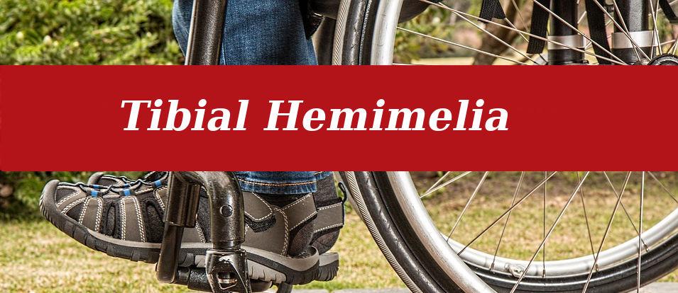
TIBIAL HEMIMELIA TREATMENT
Tibial hemimelia is a rare congenital lower limb deficiency presenting with a wide spectrum of malformation, deficiency, and duplication. It presents with a variety of presentations including severe leg & foot deformity, limb shortening, weakness in the quadriceps & absent cruciate ligaments, and patella. It can be unilateral or bilateral. Presentation in tibial hemimelia is very wide. It can present with a variety of presentations ranging from hypoplastic tibia to complete absence of tibia. The incidence of tibial hemimelia is one per million. Dr Drohr Paley has classified tibial hemimelia according to deficiency.
Type 1: hypoplastic nondeficient tibia with valgus knee joint
Type 2: dysplastic ankle with the presence of both proximal & distal physis
Type 2a: well-formed both proximal & distal tibial physis, equinovarus deformity of the foot
Type 2b: delta tibia, bracket physis, malorientation of ankle & knee joint.
Type 2c: delayed or absent ossification (part or all) of tibia with dysplastic ankle joint
Type 3: presence of knee joint, absence of distal tibial plafond.
Type 3a: absence of tibial plafond, medial and lateral
malleolus present
Type 3 b: same as type 3A with skin cleft separating tibia & fibula.
Type 4: distal tibia aplasia
Type 4a: complete absence of distal tibia from diaphysis, knee joint present.
Type 4 b: epiphysis of the proximal tibia and knee joint present but absent proximal physis
Type 5: complete tibial aplasia
Type 5a: Complete absence of tibia, patella present;
Flexion contracture of the knee, equino-varus contracture of
Dislocated foot and ankle
Type 5B: Complete absence of tibia, no patella present;
Flexion contracture of the knee, auto-centralized fibula,
Quadriceps present, knee capsule present
Type 5C: Complete absence of tibia, no patella present;
Flexion contracture of knee, dislocated fibula, quadriceps
Absent, no knee capsule present.
We follow the principle of Drohr Paley in the reconstruction of these deformities. The treatment plan is based on the classification given by Drohr Paley. In type, 1valgus deformity of the knee is corrected by hemiepiphyiodesis and limb lengthening by Illizarov. In variety with intact knee joint and tibia, length and alignment of the leg are restored in the staged manner it includes illizarov distraction to correct the deformity, tibialization of fibula, and fibula-talar arthrodesis followed by limb lengthening. In children with a complete absence of a tibia & with the presence of a patella, we use the procedure of brown arthroplasty in which the patella was utilized to make the knee joint & tibia was made use of the fibula. with the absence of patella and nonfunctioning quadriceps knee disarticulation is needed. Usually, the first surgery is done at the age of 1.5 years. Serial limb lengthening may be required. In earlier years gold standard for treatment was limb amputation with prosthetic fitting. With the door Paley approach complete reconstruction can be done in most of these children and they can have plantigrade feet and equal limb length.
Tibial hemimelia is the congenital absence of tibia. Its severity ranges from a minor deficiency in length to a complete absence of tibia. Infants can present with severe deformity & shortening of the leg, equinovarus deformity of the foot & instability of knee & ankle joints. Drohr Paley has classified this problem into five varieties based on tibial length deficiency in a progressive manner & knee problems. In mild to moderate length discrepancy lengthening of the tibia, tibialization of the fibula, and ankle fusion along with a lengthening of the tibia is required. In severe deficiency, reconstruction or fibula femoral fusion of knee joint and stabilization of ankle joint on the distal end of the fibula can be done in some cases but in a few cases, amputation & prosthesis are required. The child will need repeated lengthening and continuous monitoring. Usually, surgical reconstruction is done between the ages of 1.5 to 2 years. All these new surgical techniques are based on the concept of Drohr Paley. Trishla Foundation under the supervision of Dr. Jitendra Kumar Jain, a pediatric orthopedic surgeon has given a new lease of life in many children affected by this problem.
 Tibial Hemimelia Child with Ilizarov